Fetal hypertension
(A) Normal mid trimester umbilical cord
(B) Hypertensive term umbilical cord showing intimal injuries, medial hyperplasia, disruption of Wharton’s jelly and neovascularisation.
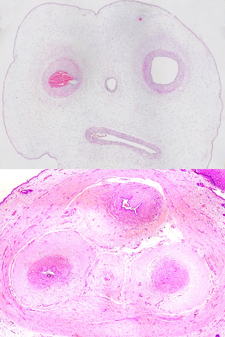
High power view of hypertensive changes in the two umbilical arteries and one umbilical vein.
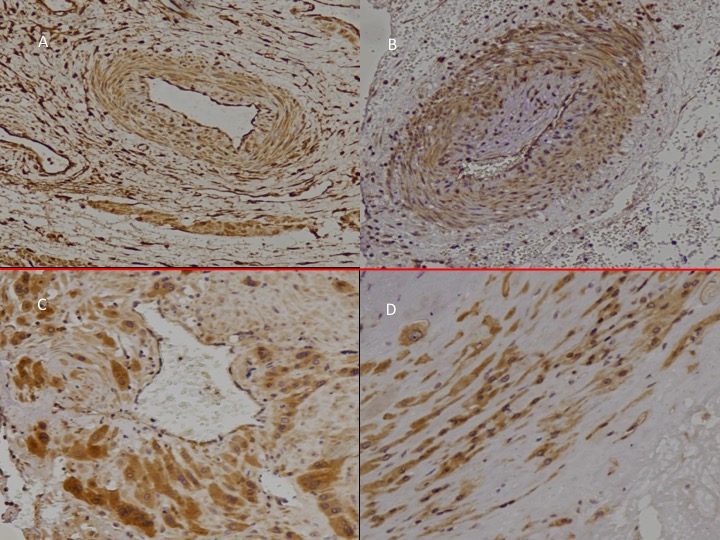
Preeclampsia
There are similar changes to the media in preeclampsia with narrowing of arterioles and purinergic P2X3 receptors expressed in thickened arteriolar walls (A-B “early-onset” PET) and myometrium (C-D,“late-onset” PET).
http://www.preeclampsiaexplained.com/wp-content/uploads/2019/05/Slide18.jpeg
Adult hypertension
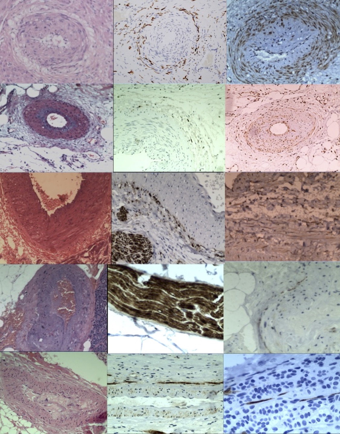
And similar changes in visceral arterioles including uterus (R1), kidney (R2), spleen (R3), pancreas (R4) and adrenals (R5) that demonstrate the origins of adrenal cortical hyperplasia (5b-c). All have common origins where neural injuries leads to release of cytokines and growth factors that cause hyperplasia of the denervated arteriolar walls.
Column 1 shows narrowed visceral arterioles stained with haematoxylin and eosin.
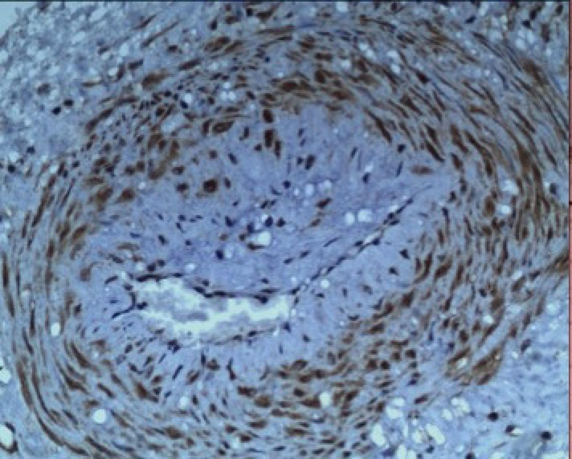
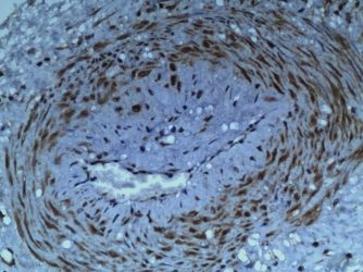
Column 2 shows proliferation of injured vasomotor nerves – note spleen in row 3, or, loss of nerves in visceral nerve bundle – note pancreas in row 4.
Column 3 shows positive, intra-arteriolar, staining (brown) for purinergic “stretch” receptors, P2X3 – but only in the uterus (row 1). The remaining viscera are negative for P2X3.